
How Fats in the Cell Membrane Keep the T Cell Receptor in Check
Background and Introduction
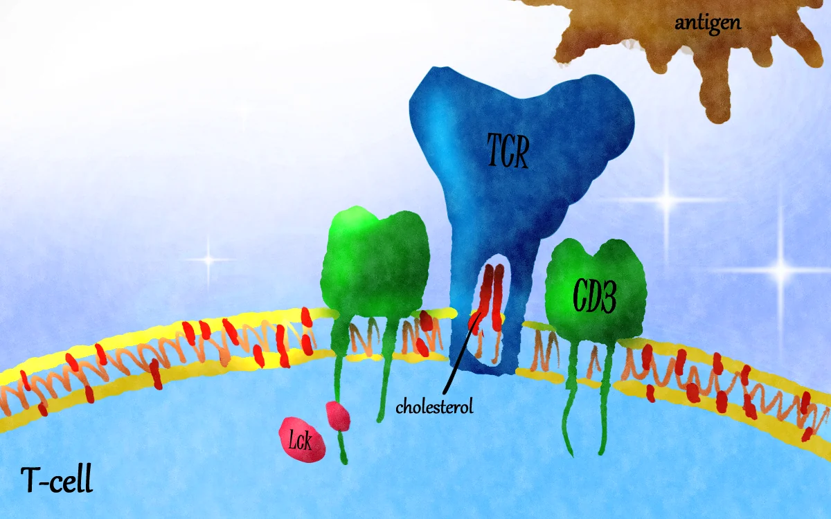
A T cell must stay quiet when there is no threat, yet respond quickly where there is one. A puzzle sits at the center of this system, where it is not fully understood how the receptor manages its active and inactive states. [4] T cells are immune cells that find and destroy infected or cancerous cells. They recognize their targets with a sensor on their surface, the T cell receptor (TCR). The TCR works with a group of partner proteins called CD3, and together they form a complex that sits in the cell’s outer membrane [1,2]. When the TCR meets its target (an antigen), the CD3 proteins relay the message inside the cell, where an enzyme called Lck attaches small chemical tags to sites on the CD3 tails. These tags switch the T cell on [1,2]. There are two main families: alpha-beta (αβ) T cells, which make up more than 90% of T cells and recognize antigen fragments displayed on other cells, and gamma-delta (γδ) T cells, which recognize a wider range of targets [1,2].
Because the receptor is embedded in a fatty membrane, researchers have long suspected that the membrane itself plays a part, particularly cholesterol [3]. This review argues that the membrane is not a passive container but part of the receptor’s on/off switch. It draws on five papers that approach the question in different ways: two that built 3D models of the receptor [2,4], one set of computer simulations [5], and two overview articles [1,3]. It first traces how the idea developed, then examines what is known about cholesterol and other membrane fats. Furthermore, this review compares what each kind of evidence can and cannot show, and finally considers what this could mean for medicine.
Key terms used in this review:
- Membrane: the fatty skin around a cell. Cholesterol is one of its main fats.
- Phosphorylation: attaching a small chemical tag (a phosphate) to a protein, which often works as an on-switch.
- Cryo-electron microscopy (cryo-EM): a method that images frozen molecules to build detailed 3D models.
- Molecular simulation: a computer model that follows how atoms move over time.
- Jurkat cells: a standard laboratory T cell line used for experiments.
- CD69: a marker that appears on a T cell’s surface when the cell has been activated.
Main Discussion
How the idea developed
The idea that cholesterol restrains the receptor is built up in stages. Earlier work, described in the papers reviewed here, shows that cholesterol binds the membrane-spanning part of the TCR and seems to hold it in an inactive shape. A related molecule, cholesterol sulfate, can take its place and also block signaling [3,4]. A 2017 overview then described cholesterol as two-sided: needed for T cell growth and for grouping receptors together, yet also able to tune how strongly these receptors signal[3]. A 2019 structure of the whole receptor gave the first detailed 3D picture but it was too blurry for the fats to be visible [4,5]. Computer simulations in 2021 placed the receptor in a realistic membrane and showed how its floppy tails behave [5]. In 2022, a sharper structure finally revealed cholesterol sitting inside the receptor [4]. In 2024, a structure of the γδ receptor also observed a cholesterol-like molecule, while showing that γδ receptors are built differently in other ways [2]. The turning point was the 2022 structure. Before it, cholesterol’s role was mostly inferred from biochemical and cell experiments; after it, there was a physical picture of how cholesterol could hold the receptor in check.
Cholesterol works like a latch on the αβ receptor
The 2022 study used cryo-EM to image the human αβ receptor and found two cholesterol molecules. They were arranged inside a tunnel formed where several parts of the receptor cross the membrane [4]. Filling the tunnel makes the receptor more compact and holds one of the signaling chains, CD3ζ (“CD3 zeta”), steady. The authors compare this to a latch that keeps a gate shut [4]. Cholesterol sulfate fits the same tunnel and grips even more tightly, which explains why it can push cholesterol out and block signaling [4].
The team then tested the latch idea. They altered the building blocks lining the tunnel so that it held less cholesterol (in the weakest case, under a quarter of the normal amount). These altered receptors became more active without any antigen: their CD3 tails were tagged more often in cells, and in laboratory T cells the altered receptor switched on activation markers more strongly [4]. Removing cholesterol from the cell surface with a protein tool called ALOD4 also boosted signaling [4]. Finally, the team imaged one of these always-on mutants and saw the CD3ζ chain tilt by about 10 degrees, shrinking the second cholesterol spot [4]. This is the first look at the receptor in an active-like shape, although a mutation, not an antigen, caused the change [4].
γδ receptors appear to use the same latch
The γδ receptor study found a cholesterol-like molecule in a similar position, although the image was less clear and only one of the two possible spots could be filled with confidence [2]. Mutants that held more cholesterol responded less to their targets, while a mutant that held less responded more [2]. As in the αβ case, removing cholesterol from the membrane increased activation [2]. The authors also noticed that the shape of CD3ζ in these variants resembled the resting and always-on forms seen in αβ receptors, and they speculate that the higher baseline activity of γδ T cells may relate to this [2]. The membrane-spanning parts of the two receptor types are also very similar, which suggests they share the same basic plan inside the membrane [2].
Cholesterol in the whole cell
At the level of the whole cell, cholesterol looks like both a friend and a brake. Activated T cells need a lot of cholesterol because they must build new membranes as they multiply. As a result, they switch on cholesterol-making machinery and switch off a sensor (called LXR) that would otherwise slow their growth [3]. Statins, drugs that block cholesterol production, hold young T cells back from maturing and dividing [3]. Cholesterol also helps pack receptors into small clusters that make them more sensitive to antigens. Blocking an enzyme that stores cholesterol (ACAT1), which raises cholesterol in the membrane, makes cancer-fighting T cells more effective [3].
These findings seem to clash with the latch model, in which removing cholesterol makes receptors more active [2,4]. The sources do not settle the matter, but they hint at reasons. The ALOD4 tool locks up cholesterol in the membrane without changing the cell’s total cholesterol [4], whereas chemicals that break up cholesterol-rich membrane patches (called rafts) calm overactive T cells in autoimmune disease [3]. One possible explanation, offered here as an interpretation and not an established fact, is that cholesterol does two separate jobs. One, it helps cluster receptors together into the same patches of the cell membrane. Two, it holds each individual receptor in its resting shape. The two effects may work against each other, so which one has the bigger impact may depend on the cell’s state.
Other membrane fats and the receptor’s floppy tails
Cholesterol is not the only fat that matters. The inner surface of the membrane carries a negative charge while the CD3 tails are rich in positive charge. In resting cells, the tails stick to the membrane, which hides their activation sites from Lck [1,4,5]. The 2021 simulations followed a complete receptor, including tails too floppy for cryo-EM to image, in a membrane modelled on the fat composition of real T cells [5]. The tails curled up and attached to the inner surface almost immediately (within about a ten-millionth of a second of simulated time). Only some activation sites poked into the membrane while others were tucked inside the curled-up tails, suggesting the sites are partly hidden when the cell is at rest [5].
The simulations also showed that the receptor attracts two rare, negatively charged fats called PIP2 and PIP3, even though together they make up only about a tenth of the inner membrane in the model. Positively charged patches in the tails, and a ring of positive charges where the receptor meets the inner membrane, drew them in [5]. The authors suggest that this crowd of charged fats may help bring Lck and related enzymes to the receptor, since those enzymes can bind such fats [5]. Cholesterol also hugged the membrane-spanning part of the receptor whether the rest of the protein was present, which fits the idea that cholesterol binding is built into the receptor’s core [5]. One overview adds that when antigen binds, charged fats around the receptor are pushed away, freeing one tail while another stays attached [1].
Two kinds of receptor, two ways of being built
Although αβ and γδ receptors share a similar membrane core, they differ in the parts that stick out of the cell. In the αβ receptor, the outer parts lock against the CD3 partners and are rigid. In the γδ receptor studied by Xin and colleagues, the outer parts do not touch the partners and are so floppy that they could not be imaged [2]. This flexibility looks finely tuned: a version with a longer linker bound its target better yet activated T cells less, suggesting that too much wobble hurts signaling [2].
Some γδ receptors also assemble as pairs. One family, called Vγ5, forms a back-to-back pair held together by a few key amino acids. Receptors with a broken pair interface still reached the cell surface but failed to bind their target or activate T cells [2]. A light-based test examining how close a pair of receptors sit suggested that Vγ2 and Vγ5 receptors form pairs while Vγ3, Vγ4, Vγ8 and Vγ9 receptors stay single, although the test cannot rule out mixtures [2]. Pairing may also hide a stretch of the receptor that other γδ receptors use to meet their targets, which would push paired receptors to rely on a different region [2].
What this could mean for medicine
Drugs that engage CD3 are already in use. Bispecific antibodies grab a T cell with one arm and a cancer cell with the other. They can also switch on any T cell regardless of its own target. While most drugs for this purpose are in trials, one named blinatumomab has been approved for some blood cancers, and others are in trials [1]. Their limits include toxic side effects, poor entry into solid tumors, and the tumor’s ability to suppress immune cells [1]. Other approaches use engineered receptors or small molecules that boost signaling [1].
The membrane findings open new options. Chen and colleagues suggest designing molecules that mimic cholesterol or cholesterol sulfate to calm overactive T cells in autoimmune disease [4]. Bietz and colleagues describe drugs that change cholesterol handling, with the direction depending on the disease: raising membrane cholesterol to boost tired tumor-fighting cells or lowering it to calm overactive ones [3]. Both approaches risk side effects, although one study found that resting T cells tolerated the loss of a key cholesterol-making switch [3]. For γδ T cells, the finding that some receptors work only as pairs suggests that assembly should be considered when designing therapies [2].
Gaps and Future Directions
The biggest missing piece is a picture of the receptor holding its target. No one has yet imaged the receptor with antigen attached, so it is still unclear how binding on the outside releases the latch on the inside [4]. Two ideas are on the table: the target may push cholesterol out directly, or it may tip the balance between a resting and an active shape the receptor flips between on its own. Current data cannot tell them apart [4]. The γδ structures also lack a bound target [2]. Most structures come from receptors removed from membranes with detergent, and none show the floppy tails [4,5], so combining target-bound structures, membrane-like settings and simulations is an obvious next step.
Several details and disagreements remain unresolved. At current image quality, cholesterol’s identity is inferred rather than directly proven [4]. The γδ images show only one confident cholesterol site compared with two in αβ [2,4], and the second site in the always-on mutant held an unidentified molecule [4]. No one has yet linked a cell’s cholesterol-handling systems [3] to how full the receptor’s cholesterol tunnel is. Researchers tested how cholesterol, charged fats, and the curled tails work together [5]. Whether cholesterol sulfate matters in real cells is unclear, since it was about 70 times less common than cholesterol in the γδ samples [2]. The simulations also require experimental checks [5].
Finally, the scope of these findings is not yet clear. Many experiments use laboratory cell lines, and only two γδ receptors were studied, so it is unknown whether pairing, linker length, and cholesterol control matter in normal γδ T cells [2]. The idea that the shape of CD3ζ explains the higher baseline activity of γδ T cells is speculation [2]. Whether drugs aimed at the cholesterol tunnel work in living animals has not been tested [4], and some CD3 proteins, such as CD3γ, have received little study [1].
Conclusion
Together, these studies suggest that the T cell receptor is not a stand-alone switch but one that is held in check by its surroundings. Cholesterol appears to latch part of the receptor in a resting position in αβ receptors and probably in γδ receptors [2,4]. Negatively charged fats and the curled-up tails help hide the activation sites and may gather signaling enzymes [5], and the cell’s overall cholesterol handling sets the wider scene [3]. The two receptor families share a membrane core but differ in how their outer parts are built [2].
This matters for two reasons. It offers a plausible answer to how a receptor can stay quiet without a threat yet respond sensitively when one appears. Although how antigen opens the latch is still unknown [4]. It also points to new treatment targets, including the cholesterol tunnel and the assembly features specific to γδ receptors, which could complement existing CD3 antibodies and cell therapies [1,2,4].
References
- Liu D, Hu X, Chen Z, Wei W, Wu Y. Key links in the physiological regulation of the immune system and disease induction: T cell receptor–CD3 complex. Biochem Pharmacol. 2024;227:116441. doi:10.1016/j.bcp.2024.116441
- Xin W, Huang B, Chi X, et al. Structures of human γδ T cell receptor–CD3 complex. Nature. 2024;630:222. doi:10.1038/s41586-024-07439-4
- Bietz A, Zhu H, Xue M, Xu C. Cholesterol metabolism in T cells. Front Immunol. 2017;8:1664. doi:10.3389/fimmu.2017.01664
- Chen Y, Zhu Y, Li X, et al. Cholesterol inhibits TCR signaling by directly restricting TCR-CD3 core tunnel motility. Mol Cell. 2022;82:1278–1287. doi:10.1016/j.molcel.2022.02.017
- Prakaash D, Cook GP, Acuto O, Kalli AC. Multi-scale simulations of the T cell receptor reveal its lipid interactions, dynamics and the arrangement of its cytoplasmic region. PLoS Comput Biol. 2021;17(7):e1009232. doi:10.1371/journal.pcbi.1009232
Spotted an error? Email [email protected] from your Wesleyan account.